Musculoskeletal
Ultrasound
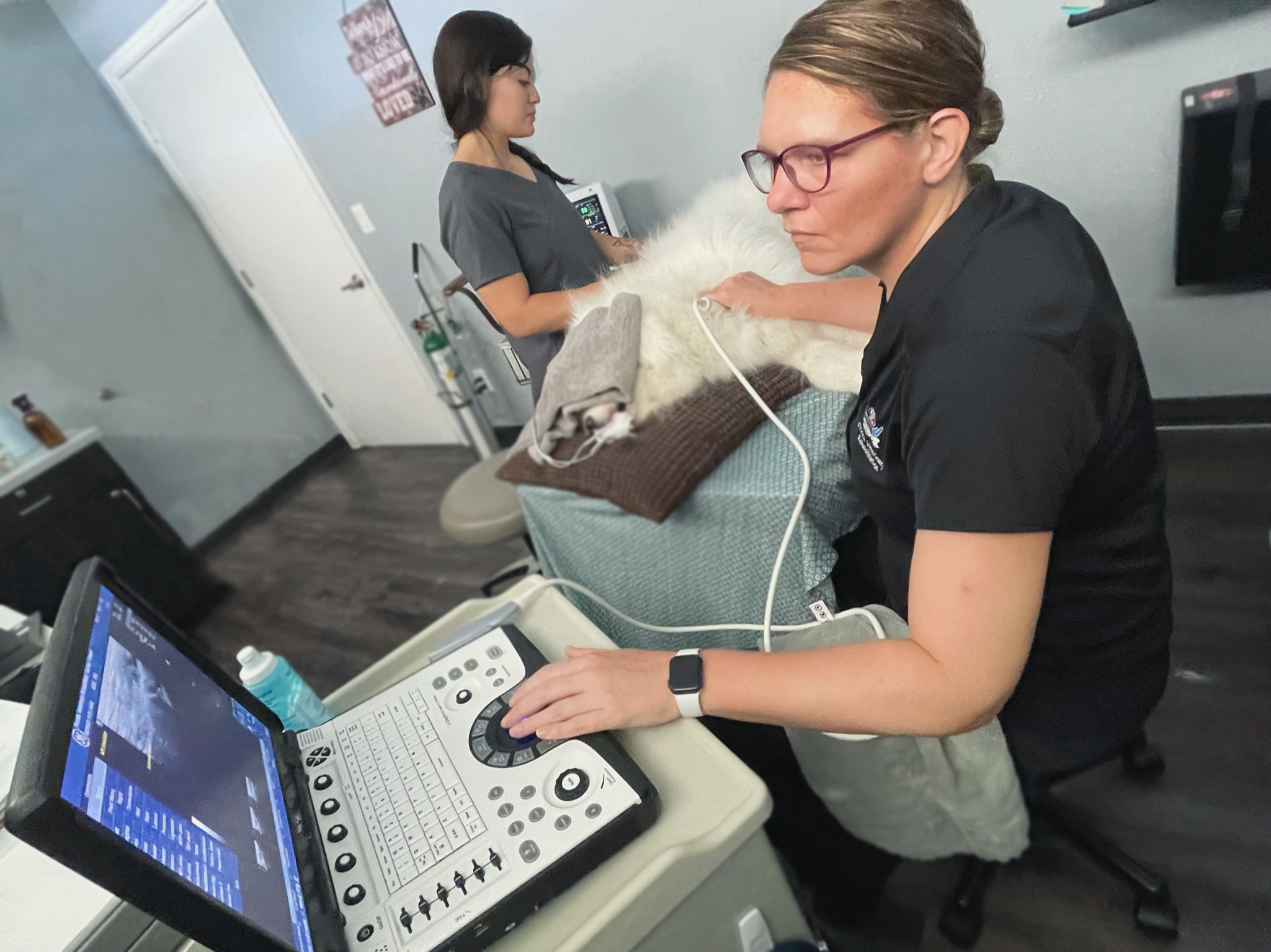
Musculoskeletal ultrasound allows detailed evaluation of your pet's tendons, ligaments, joints, and other soft tissues without radiation. This safe, non-invasive imaging technique uses high frequency sound waves to visualize structures not seen on radiographs.
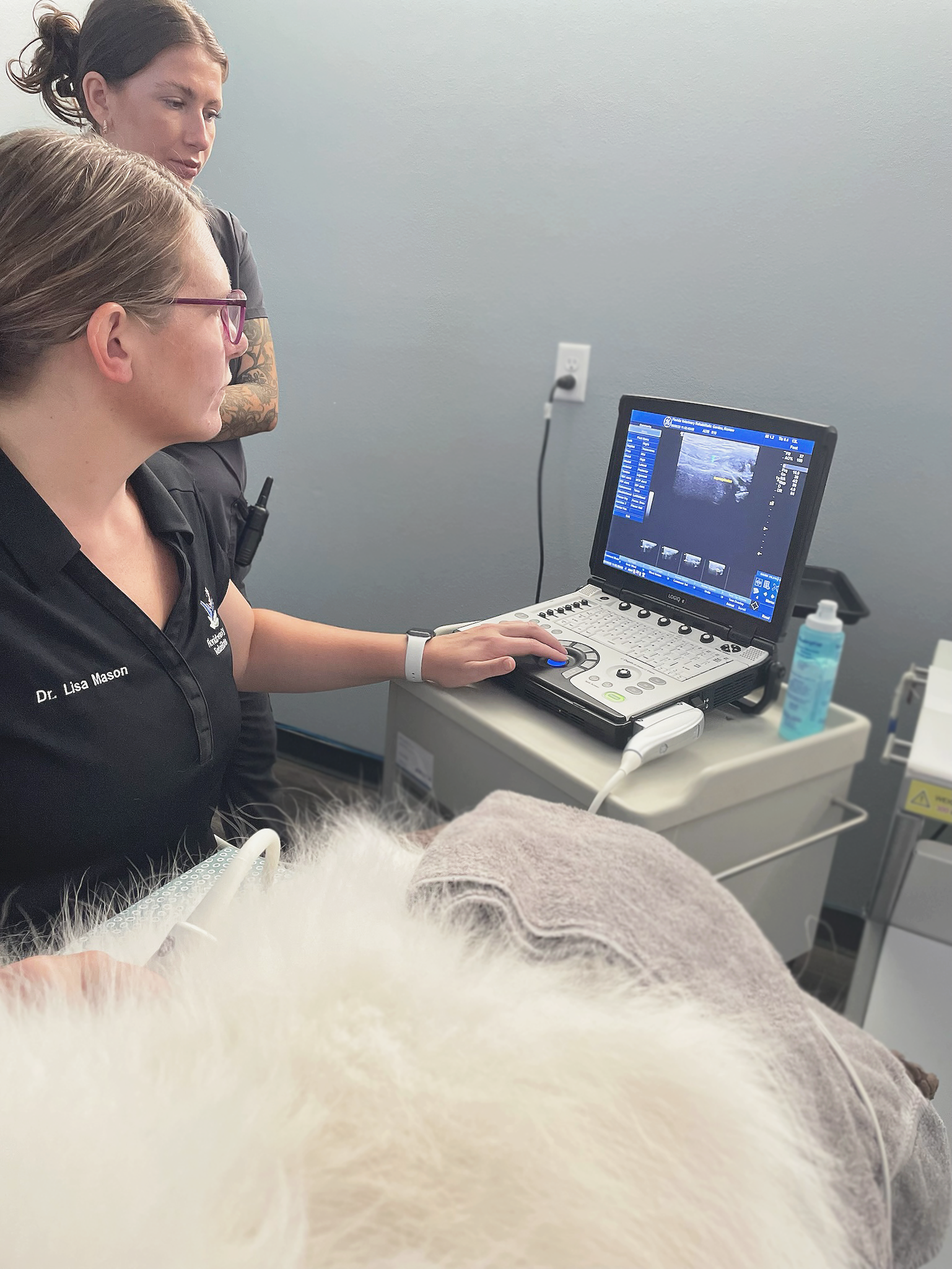
During the painless scan, a small handheld transducer is moved over the shaved area after applying gel. The transducer emits sound waves that rebound off tissues to create real-time images on the screen.
Musculoskeletal ultrasound is ideal for:
Diagnosing soft tissue injuries - tendon/ligament tears, muscle strains such as:
Biceps tendinopathy (shoulder)
Supraspinatus tendinopathy (shoulder)
Iliopsoas strain (groin)
Achilles tendon injuries/tears
Carpal and tarsal ligament injuries
Other ligament, tendon, or muscle injuries
Joint effusion and inflammation detection
Guiding joint taps and injections
Screening for injuries in canine athletes
Evaluating causes of chronic lameness
Bone surface lesions
Musculoskeletal ultrasound allows detailed evaluation and diagnosis of injuries affecting key tendons, muscles, and ligaments in the joints of dogs and cats. This imaging technique can precisely pinpoint areas of swelling, tearing, and reduced mobility to better guide our rehabilitation therapy plan.
Musculoskeletal Ultrasound FAQs
-
Musculoskeletal ultrasound uses high frequency sound waves to evaluate joints, tendons, ligaments, muscles, and related structures. It allows detailed visualization of soft tissue injury and joint abnormalities to guide rehab and treatment.
-
Ultrasound helps diagnose causes of lameness, pain, or mobility issues like arthritis, tendon/ligament tears, muscle strains, and joint instability. It provides imaging where x-rays don't give needed soft tissue detail.
-
Benefits include:
• Detailed evaluation of soft tissue structures
• Ability to compare bilateral structures
• Radiation-free imaging
• Noninvasive procedure
• Provides rehab guidance by identifying areas of injury/degeneration
• Ultrasound is also used to guide joint taps or injections and needle biopsies. It provides valuable information to customize rehab and treatment.
-
Your pet receives a gentle clipping of hair over the area. Warm gel is applied to the skin to allow the ultrasound probe to glide over the surface. Images appear on a monitor as the probe is moved. Sedation may be required for certain regions.
-
Common findings include:
• Tendon/ligament tears or inflammation
• Joint instability/effusion
• Muscle strains/hematomas
• Arthritis/intra-articular pathology
• Chronic degeneration or scarring